This article is a comparison between fNIRS vs EEG, with both being non-invasive brain imaging methods used in research and clinical settings. While fNIRS offers improved spatial resolution, EEG provides superior temporal resolution. Understanding the differences between these techniques is crucial for advancing brain imaging technology and applications.
a Table of Contents
fNIRS vs EEG: When To Use Which?
Functional near-infrared spectroscopy (fNIRS) and Electroencephalography (EEG) are two widely used non-invasive neuroimaging methods for studying real-time brain activity. While both tools serve essential roles in cognitive neuroscience and brain function monitoring, they differ significantly in their mechanisms, resolution, and ideal applications.
What is fNIRS And How Does It Work?
fNIRS monitors cerebral hemodynamic responses, which is the measurement of how blood flow in the brain shifts in response to mental tasks or stimuli, by measuring changes in oxygenated (HbO) and deoxygenated hemoglobin (HbR) using near-infrared light.
It offers better spatial resolution than EEG, particularly for surface cortical areas, and is more tolerant of movement artifacts. While fNIRS resembles functional MRI (fMRI) in principle, it is more portable and user-friendly, though its temporal resolution is slower (on the scale of seconds), as it reflects the indirect hemodynamic response to neural activity.
Understanding these foundational aspects of fNIRS is crucial for researchers evaluating its utility. For a more comprehensive overview of this powerful neuroimaging technique and its evolving applications, we invite you to explore fNIRS.
What is EEG And How Does It Work?
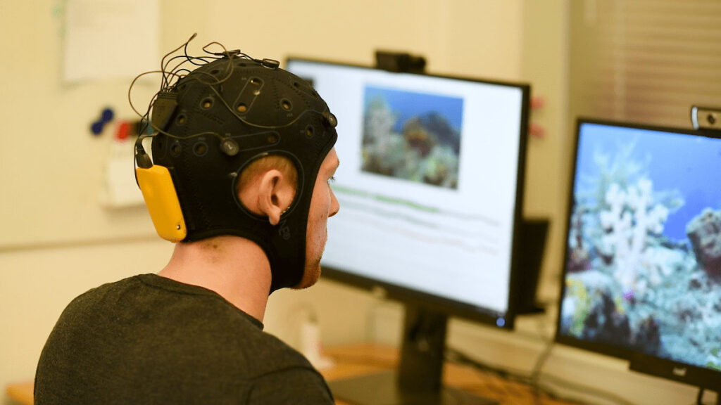
In contrast, EEG measures the brain’s electrical activity via electrodes placed on the scalp. These sensors detect voltage changes caused by synchronized firing of cortical neurons, primarily pyramidal cells. One of EEG’s greatest strengths is its exceptional temporal resolution-it captures neural dynamics on a millisecond scale.
This makes EEG ideal for analyzing rapid cognitive processes like attention, sensory perception, and motor planning. However, EEG’s spatial resolution is limited due to the dispersion of electrical signals across the skull and scalp.
fNIRS vs EEG: Where The Data Comes From
Both fNIRS and EEG are non-invasive techniques, but they differ significantly in where in the brain they measure activity-shaped by the physical properties of light and electricity as they pass through the skull.
fNIRS:
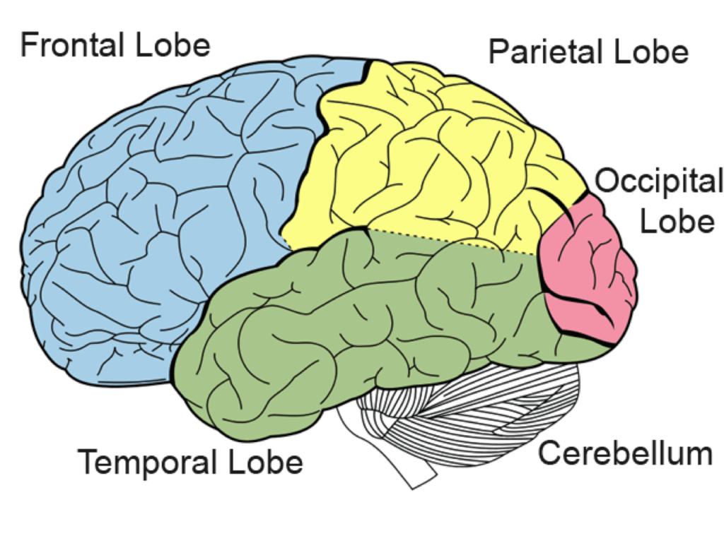
fNIRS measures brain activity in the outer layers of the cortex, primarily targeting the prefrontal and parietal regions. It detects changes in blood oxygenation just a few centimeters beneath the scalp, making it ideal for monitoring surface-level cortical areas involved in attention, emotion regulation, and higher-order cognitive processes.
EEG:
EEG captures electrical activity generated by populations of neurons in the cerebral cortex, especially from pyramidal cells aligned perpendicular to the scalp. While it records from the entire scalp, it is most sensitive to signals from superficial cortical structures and less effective at detecting activity from deeper brain regions or folded cortical areas.
Chapter: When to Use fNIRS vs EEG
Understanding when to use functional near-infrared spectroscopy (fNIRS) versus electroencephalography (EEG) is essential for designing effective and impactful cognitive neuroscience experiments. While both are non-invasive neuroimaging techniques that offer valuable insights into brain function, they measure different physiological processes, operate on different temporal and spatial scales, and are suited to distinct research contexts.
1. Nature of Brain Activity Being Studied
The primary distinction lies in the physiological signal each modality captures. EEG measures electrical potentials generated by synchronized neuronal activity, offering a direct view of neural dynamics. fNIRS, by contrast, measures changes in blood oxygenation associated with neural activity-an indirect marker relying on neurovascular coupling.
- Use EEG when your study demands high temporal resolution, such as capturing rapid cognitive processes like stimulus perception, language processing, or decision onset.
- Use fNIRS when your focus is on sustained cognitive states, workload, or affective processing localized in the cortex, particularly the prefrontal region.
2. Temporal vs Spatial Resolution Needs
EEG offers millisecond-level temporal resolution but limited spatial accuracy due to the conductive properties of the skull and scalp. fNIRS offers superior spatial resolution over surface cortical areas but is constrained by the delay of the hemodynamic response (2–6 seconds).
- Choose EEG for tasks requiring precision timing-e.g., ERP studies, rapid sequence tasks, or brain-computer interfaces (BCIs).
- Choose fNIRS for spatial mapping of brain regions involved in sustained attention, problem-solving, or emotional engagement, especially when frontal cortical activity is of interest.
3. Experimental Environment and Movement Tolerance
fNIRS systems are relatively robust to movement artifacts and more portable than traditional EEG setups, making them ideal for field studies, studies involving children, or studies requiring ambulatory participants.
- EEG is better suited for highly controlled lab environments.
- fNIRS is preferable in mobile or real-world contexts (e.g., classroom settings, sports performance, or driving simulations).
4. Multimodal Opportunities
For comprehensive cognitive or affective neuroscience investigations, integrating both EEG and fNIRS offers a hybrid approach-providing high temporal and spatial fidelity. This is especially useful in studies aiming to model neurovascular coupling or decode complex mental states in real time.
Conclusion:
The choice between EEG and fNIRS should be guided by the research question, the cognitive processes under investigation, the required resolution (temporal vs spatial), and the logistical constraints of the study population or environment. In many cases, a combined EEG-fNIRS approach yields the most comprehensive insight into brain function..
Key Considerations for Simultaneous EEG + fNIRS Use:
- Sensor Placement Compatibility
- Both systems often use the international 10–20 system for electrode/sensor placement. To avoid interference:
- Use high-density EEG caps with pre-defined fNIRS-compatible openings.
- Some fNIRS systems are designed to be embedded within EEG caps or mounted using optode holders that avoid electrode contact points.
- Use high-density EEG caps with pre-defined fNIRS-compatible openings.
- Both systems often use the international 10–20 system for electrode/sensor placement. To avoid interference:
- Hardware Integration
- Some vendors offer integrated systems, while others require synchronization via triggers or shared clock systems.
- EEG and fNIRS can typically be synchronized using external hardware (e.g., TTL pulses or parallel ports) or shared acquisition software.
- Some vendors offer integrated systems, while others require synchronization via triggers or shared clock systems.
- Motion and Signal Artifacts
- While fNIRS is more motion-tolerant, adding EEG increases complexity and sensitivity to movement artifacts.
- To minimize artifacts:
- Use tight but comfortable cap fittings.
- Avoid overlapping sensors.
- Employ motion correction algorithms during preprocessing.
- Use tight but comfortable cap fittings.
- While fNIRS is more motion-tolerant, adding EEG increases complexity and sensitivity to movement artifacts.
- Data Fusion Considerations
- EEG and fNIRS capture fundamentally different signals (electrical vs hemodynamic), so separate preprocessing pipelines are needed before integration.
- Data fusion techniques include joint Independent Component Analysis (jICA), canonical correlation analysis (CCA), and machine learning approaches that combine feature sets from both modalities.
- EEG and fNIRS capture fundamentally different signals (electrical vs hemodynamic), so separate preprocessing pipelines are needed before integration.
When to Use Separate Studies Instead:
- If sensor interference is unavoidable due to head size or equipment constraints, such as VR studies.
- If the task involves excessive movement (e.g., walking), which may degrade EEG quality.
Conclusion: Making the Right Choice in the fNIRS vs EEG Debate
When it comes to designing neuroscience or human factors studies, the fNIRS vs EEG decision ultimately hinges on your specific research goals. If you’re aiming to capture fast, transient brain responses with millisecond precision, EEG remains the gold standard. On the other hand, if your focus is on localized cortical activity during sustained cognitive or emotional states-especially in real-world settings-fNIRS may be the more suitable choice.
In some cases, the best answer to fNIRS vs EEG is not either/or but both. A combined approach allows researchers to harness the strengths of each modality, delivering richer, multidimensional insights into the brain’s electrical and hemodynamic responses. Whether used separately or together, selecting the right method (or combination) ensures your research is both scientifically rigorous and practically impactful.
Comparison Sheet Between fNIRS vs EEG:
| Feature | EEG (Electroencephalography) | fNIRS (Functional Near-Infrared Spectroscopy) |
What It Measures | Electrical activity of neurons | Hemodynamic response (blood oxygenation levels) |
Signal Source | Postsynaptic potentials in cortical neurons | Changes in oxygenated and deoxygenated hemoglobin |
Temporal Resolution | High (milliseconds) | Low (seconds) |
Spatial Resolution | Low (centimeter-level) | Moderate (better than EEG, but limited to cortex) |
Depth of Measurement | Cortical surface | Outer cortex (~1–2.5 cm deep) |
Sensitivity to Motion | High – susceptible to movement artifacts | Low – more tolerant to subject movement |
Portability | High – lightweight and wireless systems available | High – often used in mobile and wearable formats |
| Setup Complexity | Moderate – requires electrode gel and scalp prep | Moderate – optode placement with minimal skin contact |
| Cost | Generally lower | Generally higher, especially with high-density systems |
Best Use Cases | Fast cognitive tasks, ERP studies, sleep research | Naturalistic studies, child development, motor rehab |
Multimodal Use | Often combined with fNIRS, fMRI, or eye tracking | Commonly integrated with EEG, motion tracking, etc. |
Free 59-page EEG Guide
For Beginners and Intermediates
- Get a thorough understanding of the essentials
- Valuable EEG research insight
- Learn how to take your research to the next level