Neuropsychology studies how the brain drives behavior, linking neurology and psychology. From ancient Egyptian head injury records to modern EEG, fMRI, and eye tracking, it reveals how brain regions shape memory, cognition, and action. Landmark cases like H.M. showed the hippocampus’ role in memory, while modern research maps atypical brain activity in autism, schizophrenia, and other conditions.
Table of Contents
What is neuropsychology exactly? About 3500 years ago, an Egyptian physician marked down with a reed pen a list of ailments on papyrus, and gave a diagnostic opinion for each. Treatments were either treatable, could be contended with, or were likely incurable. 14 of the 48 cases were head injuries – the earliest documented cases of damage to the brain [1]. This was, in some senses, the beginning of neuropsychology.
Neuropsychology is the study of how the brain affects behavior. It draws a line between neurology and psychology and investigates how these processes interact. When the ancient Egyptian physician remarked that a cut to the head should be examined by testing the patient’s ability to look down, they were making a direct link between the action of the brain and the action of the body. This approach essentially underlines all of modern neuropsychology.
Today, while we have moved on from reed pens and papyrus, we still consider how the brain impacts our behavior. We also now have better tools to make these studies. Modern imaging methods have taken us from feeling wounds with our fingers to recording the pulsing of blood in the brain and its electrical activity, without ever having to touch the patient. Non-invasive methods have given access to an understanding of the brain and behavior like never before.
Below, we will define the field of neuropsychology, explain its recent origins, and discuss the modern science of neuropsychology.
What is Neuropsychology?
Neuropsychology takes the approach that the activity of the brain determines the cognitive or behavioral output of the body. The field officially started in 1963 with the launch of the scientific journal Neuropsychologia [2]. Although the approach of neuropsychology, and even the word itself have origins at other moments in time, the journal finally and firmly established the topic as a scientific field of study.
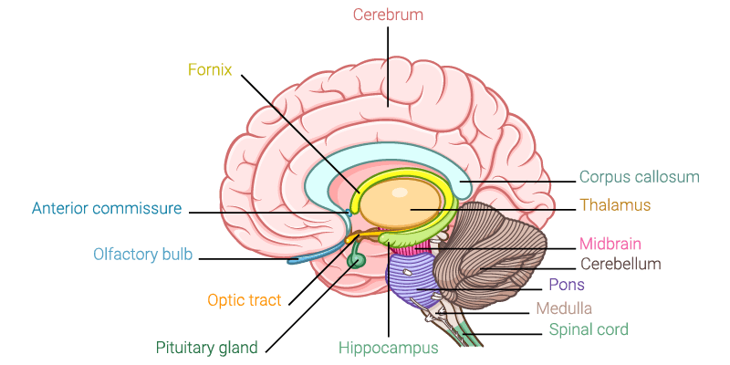
It has historically used examples of traumatic brain injuries to determine which part of the brain does what. By examining a patient with damage to (for example) the hippocampus, and discovering that the patient’s memory encoding abilities are also damaged, it’s possible to make a link between the hippocampus and memory encoding.
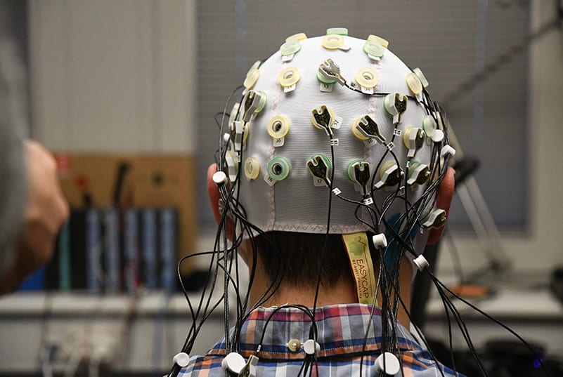
With the rise of brain imaging methods, such as EEG and fMRI, it has also been possible to study brain processes and their relationship to behavior. These methods largely forego the need for lesion studies, as brain processes can be examined in a rigorous and controlled manner. Below we will discuss some of these breakthroughs in more detail.
As a result of these brain lesion, and brain imaging studies, we today know that distinct areas of the brain are responsible for distinct tasks. This is however a fuzzy definition – brain activation is rarely related exclusively to a single specific action. There are however enough commonalities in how brain areas are associated with certain actions that we can reliably point to a distinct neural area and say which processes it is usually involved in.
Neuropsychology Research
One of the most pivotal studies in neuropsychology came about as a result of an accident. Henry Molaison contracted severe epilepsy from a young age (possibly due to a fall from a bike), which became worse later in life. At the age of 27, the seizures became so frequent and so severe that keeping a job became impossible.
As a last resort measure, neurosurgery was planned. The premise was relatively simple (insofar as neurosurgery can be) – remove the areas of the brain that are triggering the seizures. What wasn’t known at the time (yet very quickly found out) was that one of the areas – the hippocampus – is crucial for memory formation. Molaison awoke from the surgery and was essentially never able to form new memories again [5].
The dramatic impact on brain function led to the case (known by the patient’s initials, H.M.) being one of the most influential neuropsychology studies. Work by Brenda Milner and others helped uncover crucial facets of hippocampal function and its association with memory formation. While we now know that the relationship between memory and the hippocampus isn’t one-to-one [6], these initial findings were instrumental for understanding how the brain forgets and remembers.
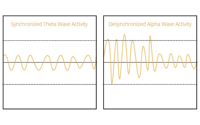
One of the most highly cited articles within neuropsychology, and neuroscience more broadly [3], concerns the relationship between brain activity as recorded by EEG, and memory formation [4]. Wolfgang Klimesch’s review of the relationship between alpha and theta oscillations to cognition and memory (with 5314 citations), consolidates the findings of several pivotal pieces of neuropsychological research.
Klimesch posits that patterns of brain activity shift reliably when new information is encoded or when semantic long-term memories are retrieved (you can read some of the details of the article in this blog post). The article exemplifies the neuropsychological approach of relating brain to function.
A less conventional neuropsychological study, also using EEG, was carried out in 1983 with dramatic findings. Using just a timer and a buzzer, Benjamin Libet recorded EEG activity that appeared to undermine the very concept of consciousness.
By asking participants to tap their finger while noting down when the urge to do so arose, Libet detected brain activity that appeared to show that the conscious choice to act was preceded by increased brain activity. In other words – the brain decided to move before the participant. The concept of free will appeared to be on shaky ground.
It took thirty years before this study was truly challenged, when Aaron Schurger found that the signals that arise before the conscious awareness of an action also occur spontaneously – even when there is no task at hand. The signals did however differentiate when the action was consciously carried out. This suggested that the signals need to pass a threshold of awareness to actually be acted upon. Free will appears to be saved, although only on the basis that it’s controlled by randomly-generated neural activity – maybe not as encouraging as first thought. The debate continues.
Beyond EEG
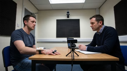
While neuropsychology has classically used direct brain imaging methods, more recent approaches have also utilized other techniques. Researchers have used tools such as eye trackers to quantitatively examine behavioral processes.
For example, initial research using eye tracking to measure the eye movements of people with schizophrenia suggested that the smooth pursuit process was dysfunctional [7]. Later studies explored this in more depth, finding that general frontal lobe dysfunction rather than schizophrenia per se is likely behind the findings [8, 9].
Another prevalent example of relating brain to behavior with the help of eye tracking has been through studies investigating autism. On a group level, people with autism have been shown to fixate less on the eye region of characters within movies [10]. Other research has shown that autism was more likely to develop later in life for 6-month old babies who exhibited a reduced overall viewing time of the presented social scenes [11]. These results suggest that behavioral outcomes such as those tested by eye trackers can inform an understanding of atypical brain processes.
Findings from other biosensors have also shown how the activity of the brain can be related to physical output. For example, damage to the right, but not left hemisphere tends to reduce or abolish electrodermal activity [12], showing a link between the right hemisphere and the sympathetic nervous system (the activity of which is reflected in electrodermal activity).
Further research has shown that an area of the brain known as the ventromedial frontal region may be particularly important for modulating skin conductance responses to psychological, but not physiological stimuli [13], illustrating the connection between all of the components within neuropsychology – the mind, body, and brain.
Conclusion
The field of neuropsychology, while in its relative infancy, has been central to our understanding of behavior – why humans act the way they do. By developing an understanding of the brain through behavior, a framework has been formed by which the complex neurological processes that form who we are can be set and built upon.
A range of methods have been central to forming this understanding, from the crude and unpredictable nature of lesion studies, to brain imaging and beyond. Each has developed ideas surrounding the way in which we understand the brain.
If you’d like to learn more about human behavior, including from other theoretical approaches, download our free guide below.
Free 52-page Human Behavior Guide
For Beginners and Intermediates
- Get accessible and comprehensive walkthrough
- Valuable human behavior research insight
- Learn how to take your research to the next level

References
[1] Sanchez, G.M., Burridge, A.L. (2007). Decision making in head injury management in the Edwin Smith papyrus. Neurosurg Focus 23(1):E5
[2] Berlucchi, G. (2010). “Neuropsychology: theoretical basis” in Encyclopedia of neuroscience. 1001–1006. 10.1016/B978-008045046-9.00996-7
[3] Yeung, A., Goto, T. K., & Leung, W. K. (2017). At the Leading Front of Neuroscience: A Bibliometric Study of the 100 Most-Cited Articles. Frontiers in human neuroscience, 11, 363. doi:10.3389/fnhum.2017.00363
[4] Klimesch W. EEG alpha and theta oscillations reflect cognitive and memory performance: a review and analysis. Brain research. Brain research reviews. 1999;29:169–195. doi: 10.1016/S0165-0173(98)00056-3.
[5] .
[6] Diana, R.A., Yonelinas, A.P., Ranganath, C. (2007). Imaging recollection and familiarity in the medial temporal lobe: a three-component model. Trends in Cognitive Sciences. ;11:379–386. doi: 10.1016/j.tics.2007.08.001.
[7] Holzman, P. S. (1975). Smooth-pursuit eye movements in schizophrenia: Recent findings. In D. X. Freedman (Ed.), Biology of the major psychoses (pp. 217-231). New York: Raven Press.
[8] Clinical, Neuropsychological, and Brain Structural Correlates of Smooth-Pursuit Eye Tracking Performance in Chronic Schizophrenia
[9] Holzman, P.S. Levy, D.L., Proctor, L.R. (1976). Smooth pursuit eye movements, attention, and schizophrenia. Archives of General Psychiatry, 45:641-647.
[10] Klin, A., Jones, W., Schultz, R., Volkmar, F., and Cohen, D. (2002). Visual fixation patterns during viewing of naturalistic social situations as predictors of social competence in individuals with autism. Arch Gen Psychiatry, Sep;59(9):809-16.
[11] Shic, F., Macari, S., and Chawarska, K. (2014). Speech Disturbs Face Scanning in 6-Month Olds who Develop Autism Spectrum Disorder. Biol Psychiatry, Feb 1; 75(3): 10.1016.
[12] Tranel, D. and Hyman, T. (1990). Neuropsychological correlates of bilateral amygdala damage. Archives of Neurology, 47, 349–55.
[13] Lane, R. D., & Nadel, L. (Eds.). (2000). Cognitive neuroscience of emotion. New York: Oxford University Press.