Unravel the distinctions between EEG, MRI, and fMRI – three pivotal brain imaging techniques. Understand how each method works, their unique advantages, and applications in neuroscience research.
Table of Contents
Understanding human thought and behavior can take many approaches, but to really understand how the brain works, you need to look inside it. This needn’t be as gruesome as it sounds, as many brain imaging methods today are entirely noninvasive.
Below we will go through the most common brain imaging techniques – EEG, and (f)MRI, to see how they work, and how they compare, looking at the advantages and disadvantages of each. Reading this article will enable readers to get a better understanding of EEG, MRI, and fMRI and how they can be used to further our understanding of the brain and behavior.
What is EEG?
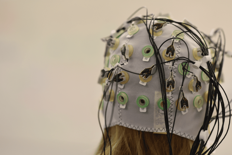
EEG (electroencephalography) measures the electrical activity of our brain via electrodes that are placed on the scalp. It tells us, from the surface measurements, how active the brain is.
This can be useful for quickly determining how brain activity can change in response to stimuli, and can also be useful for measuring abnormal activity, such as with epilepsy [1].
How does EEG work?
The brain is an electrical system – all of our thoughts (conscious or otherwise) are generated through a network of neurons, that send signals to each other with the help of electrical currents. The more electrical signals, the more neuronal communication, which corresponds to more brain activity.
The electrodes of an EEG headset can’t detect changes in single neurons, but instead detect the electrical changes of thousands of neurons signalling at the same time.
The signal from the electrodes is then sent to an amplifier, that (no surprises here) amplifies the signal. A computer then receives this signal, and can generate various maps of brain activity, with a rapid temporal resolution.
A drawback for EEG is the spatial resolution – as the electrodes measure electrical activity at the surface of the brain, it is difficult to know whether the signal was produced near the surface (in the cortex) or from a deeper region.
There are calculations that can be applied that attempt to get around this limitation (e.g. [2]), but it remains a challenge for EEG research.
Check out: What is EEG and How does it Work
What is MRI?
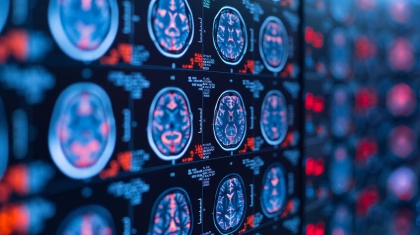
MRI (magnetic resonance imaging) provides a map of the brain – how it looks at a set moment in time.
This structural information can be useful for determining how the sizes of certain brain areas compare across people, or if there is something abnormal about a particular brain (a tumor for example).
How does MRI work?
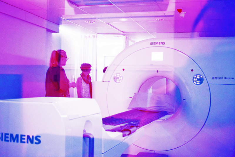
MRI is a complex imaging methodology, but we’ll try to give you an overview here.
As the name suggests, magnets are central to magnetic resonance imaging, but quite a bit stronger – roughly 1,000 to 3,000 times stronger than the average fridge magnet.
The magnetic field from the MRI interacts with the protons in our hydrogen atoms [3] (it’s of course pretty handy that we are 70% water – there are plenty of hydrogen atoms for the magnet to affect).
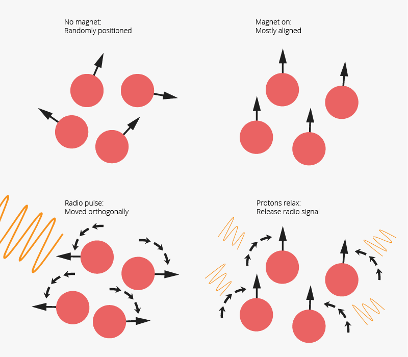
Usually, these protons are facing in random directions, but the magnetic field makes a significant portion of them align in the same direction. So, we’re lying in the MRI machine, and the protons in the hydrogen atoms (that are in the water in our body), are mostly pointing the same way. Phew.
For the next step, a radio pulse is emitted (just like a normal radio signal, just much quicker). This also interacts with the protons, essentially turning them to the side. But, as the radio frequency only happens for a moment, the protons relax back to their aligned state before.
This is the crucial bit – as the protons relax, energy is released which can be detected by sensors in the MRI machine. Through some calculations (that are beyond the scope of this blog post, but see here: [4]), the computer can determine what the tissue looked like, depending on this energy that is released, and show us an image of the tissue.
Of course, MRI only shows us a static image of the brain – an anatomical image, not of the brain’s actual activity. So how can we get an image of the brain activity? This is where fMRI comes in.
What about fMRI?
If I want to move my right arm, a few things need to happen. A certain part of my brain will increase its activity to send the message to complete this action, and that area of the brain will receive ever-so-slightly more oxygen-rich blood.
For fMRI, the same things happen as with MRI – the energy emitted from the relaxation of protons is measured – but the calculations are instead aimed at determining how the amount of oxygenated blood flow changes.
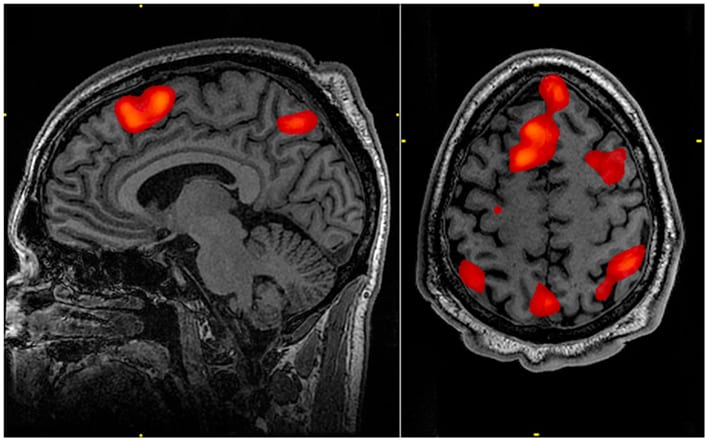
If there is more oxygenated blood in one part of the brain compared to others, then chances are that this brain area is more active [5]. This is known as the Blood-Oxygenation Level Dependent response (otherwise known as BOLD).
This is the data that we see with fMRI, often visualized over an MRI image.
One drawback with fMRI is the temporal resolution. As it takes several seconds for the blood flow to change, and the actual recording is limited by computational factors, the data collection is slowed down.
This often means that participants are exposed to a stimuli multiple times, and different timepoints of their brain response are recorded each time (e.g. the response is recorded at stimuli onset the first time, 10ms after stimulus onset the second time, and so on) [6].
This can of course undermine the accuracy of recording a novel response, but does provide a full range of brain responses.
Interested in Human Behavior and Psychology?
Sign up to our newsletter to get the latest articles and research send to you
How do they all compare?
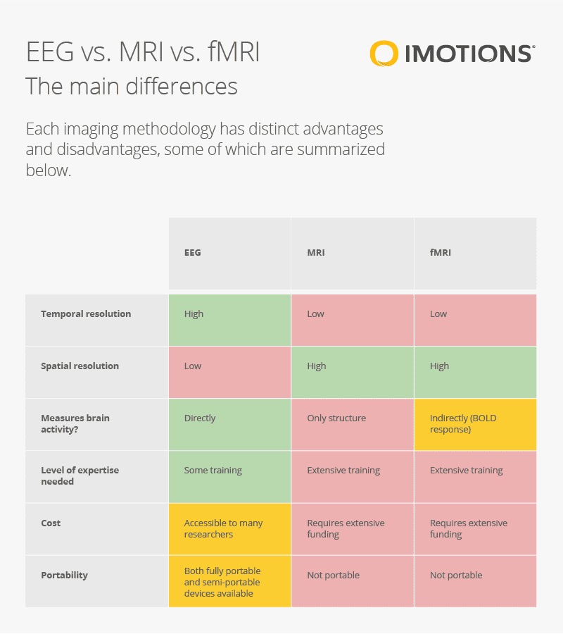
As we’ve learnt above, there are several differences in how the brain imaging information is provided by each technology.
There are also additional things to consider – the cost of an MRI machine is considerably higher than an EEG (both for purchase and maintenance), and the level of training required is much more extensive.
Doing field work with MRI / fMRI also isn’t going to happen, as there’s no way to make such a machine truly portable.
Setting up an experiment with EEG can also be done without too much hassle – sometimes as easy as placing a headset on, and checking the data quality. Metrics that are automatically calculated can also provide quick insights about human behavior with EEG.
While climbing into an MRI machine can be completed easily enough, deciding on which radio pulse to deliver, or analyzing the data is a task that requires a high level of knowledge and expertise.
We’ve put together the advantages and disadvantages of each in the table below.
Which should you use?
As always, this depends on your research question. If you are more concerned with structural and functional detail, then MRI or fMRI could well be your choice if you are able to make the considerable investment required.
For quicker, affordable, and accessible insights about brain function, with a tight temporal resolution, EEG is the method of choice.
If you’d like more guidance in deciding on the method of choice for your research, then reach out and talk to our team.
I hope you’ve found this discussion and comparison of MRI, fMRI, and EEG helpful. If you’d like to get an even deeper understanding of EEG, then download our free guide below!
Free 59-page EEG Guide
For Beginners and Intermediates
- Get a thorough understanding of the essentials
- Valuable EEG research insight
- Learn how to take your research to the next level

References
[1] Noachtar, S., & Rémi, J. (2009). The role of EEG in epilepsy: A critical review. Epilepsy & Behavior, 15(1), 22-33. doi: 10.1016/j.yebeh.2009.02.035
[2] Oja, E., Harmeling, S., & Almeida, L. (2004). Independent component analysis and beyond. Signal Processing, 84(2), 215-216. doi: 10.1016/j.sigpro.2003.11.005
[3] Mills, A., Sakai, O., Anderson, S., & Jara, H. (2017). Principles of Quantitative MR Imaging with Illustrated Review of Applicable Modular Pulse Diagrams. Radiographics, 37(7), 2083-2105. doi: 10.1148/rg.2017160099
[4] Jung, B., & Weigel, M. (2013). Spin echo magnetic resonance imaging. Journal Of Magnetic Resonance Imaging, 37(4), 805-817. doi: 10.1002/jmri.24068
[5] Hillman, E. (2014). Coupling Mechanism and Significance of the BOLD Signal: A Status Report. Annual Review Of Neuroscience, 37(1), 161-181. doi: 10.1146/annurev-neuro-071013-014111
[6] Hennig, J., Speck, O., Koch, M., & Weiller, C. (2003). Functional magnetic resonance imaging: A review of methodological aspects and clinical applications. Journal Of Magnetic Resonance Imaging, 18(1), 1-15. doi: 10.1002/jmri.10330