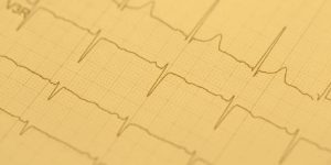
ECG (electrocardiography) is a method of collecting electrical signals generated by the heart. This allows us to understand the level of physiological arousal that someone is experiencing, but it can also be used to better understand someone’s psychological state.
Below we’ll go through the importance of physiological arousal in emotions, the physiology of the heart, how the activity can be measured, and what parameters are of interest.
- Physiology and function of the heart
- Emotional Arousal
- How to measure heart activity?
- Cardiac parameters of interest
- Why combine ECG with other sensors?
- Download our FREE Guide to ECG
Physiology and function of the heart
Before we dig deeper into the fundamentals of ECG, let’s go through the basics of heart physiology and function:
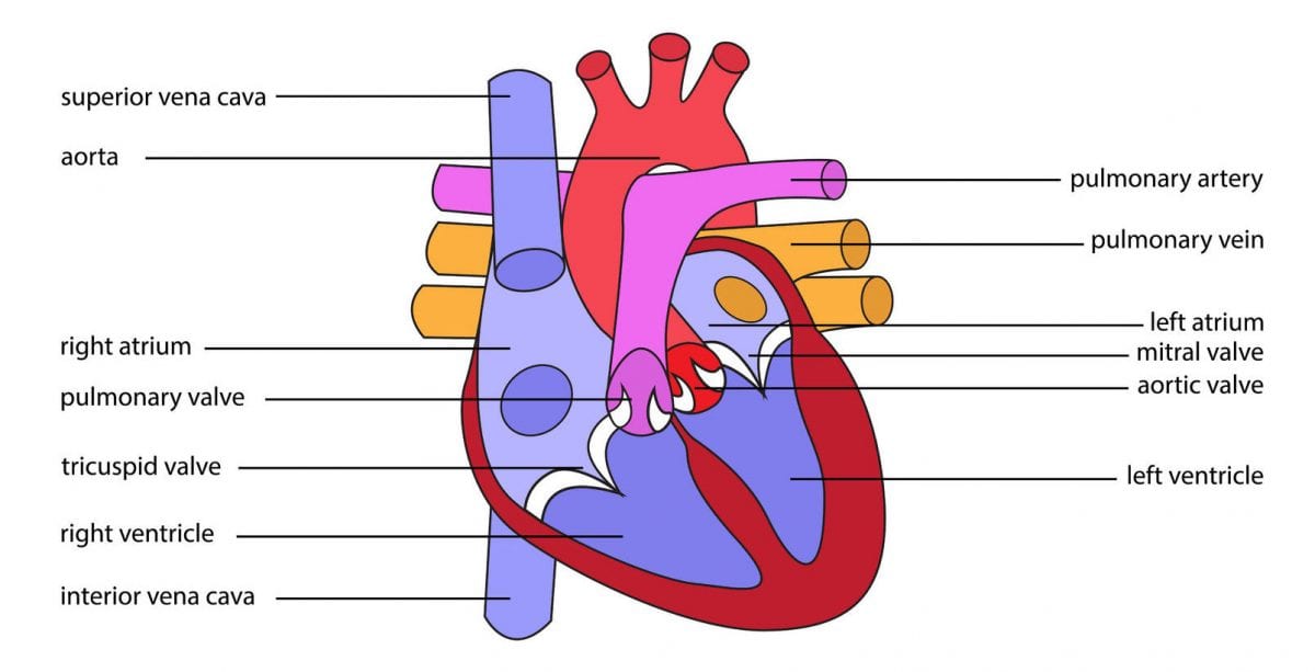
- The heart has four chambers. The upper two chambers (the left and right atrium) are entry points into the heart, while the lower two chambers (the left and right ventricle) are contraction chambers that send blood out to the body.
- The blood comes into the top atria after returning from either the lungs or other parts of the body and is then pumped into the lower ventricles, before being pumped back out again (to the alternate destination to where it just was).
- The circulation of the heart is therefore split into a “loop” – one part goes through through the lungs (the pulmonary loop) and the other through the body (the systemic loop).
- These loops ensure that the blood is either being oxygenated in the lungs or is providing that oxygen to other parts of the body where it is needed.
- The cardiac cycle refers to a complete heartbeat from its initial squeezing of the blood into the atria, to the emptying of the ventricles. The frequency of the cardiac cycle is reflected as heart rate (beats per minute, or bpm).
- The heart operates automatically – it is self-exciting (this is a unique feature compared to other muscles in the body that require nervous stimuli for excitation).
- The rhythmic contractions of the heart occur spontaneously, but are sensitive to nervous or hormonal influences, particularly to sympathetic (arousing) and parasympathetic (decelerating) activity.
Emotional Arousal
The heart doesn’t act alone. While it can operate independently, it has many deep and intricate connections to the brain, which can set its rhythm depending on the needs of the body at any given moment. The activity of the heart can also interact with the brain, and ultimately change how we feel.
An influential study by Schacter and Singer in 1962[1] showed how this occurs. Participants were told that their vision would be tested after they were given vitamins – it appeared to the participants to be a fairly basic medical study. They were instead either given epinephrine (also known as adrenaline), or a placebo drug.
They were then told that the effects of the drug were the same as epinephrine, or that they might feel some discomfort, or they were told nothing at all.
Epinephrine is known to activate the central nervous system, increasing the heart rate, causing pupil dilation, and creating an overall more activated state (if you’ve ever had an “adrenaline rush” then you know what the effects are like). The participants were then placed in a waiting room with a person who also appeared to be waiting. This person was however an actor employed by the researchers, who pretended to be angry or happy.

The angry or happy conditions were routines that the actor performed (for example, step fifteen of the happy condition reads: “Stooge [the actor] replaces the hula hoop and sits down with his feet on the table. Shortly thereafter the experimenter returns to the room.”) that were intended to influence the emotional state of the participant.
The participant then had to complete a questionnaire about how they were feeling. They found that the emotional states of the participants were influenced by the actions of the actor – those who were in the happy or angry condition reported being happier or angrier than those in the control conditions. They also experienced that emotion more strongly after they’d received a dose of epinephrine.
But there was a twist – the participants rarely pointed towards the drug, or the actor’s performance as influencing how they felt, instead claiming that they were just having a good or bad day, and that it had nothing to do with the experiment. This suggested that participants (and people more generally) are less aware of the malleability of their emotional states, misattributing how they are influenced.
Why do we bring that up? This experiment, and many others since, show how physiological arousal can be related to emotional arousal, yet the reasons for our emotional state can be more difficult to know.
With this in mind, monitoring physiological or emotional arousal using biosensors presents an objective alternative to the otherwise subjective inferences that individuals inevitably make. Heart activity is closely linked to arousal, making it a great tool for helping to understand mental states in more detail.
But how can the beat of your heart be recorded and made available for analysis and interpretation? And what do the results mean? Let’s figure this out.
How to measure heart activity?
Heart activity can be recorded in two ways:
1. Electrocardiography (ECG, EKG)
ECG records the electrical activity generated by heart muscle depolarizations (a negative change in the electric charge), which propagates as pulsating electrical waves towards the skin. Although the amount of electricity is in fact very small, it can be picked up reliably with ECG electrodes attached to the skin (in microvolts, or uV).
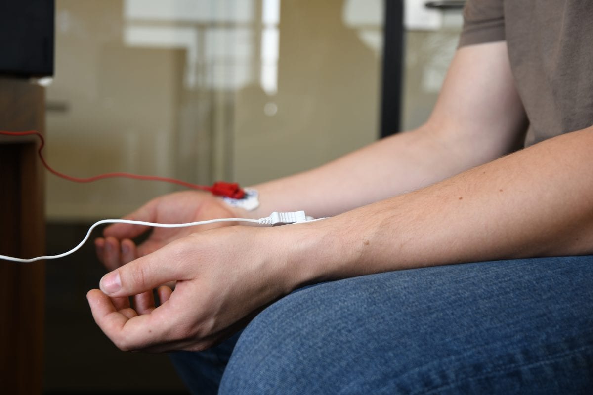
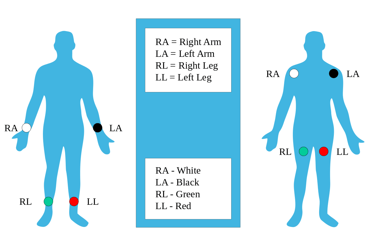
The full ECG setup comprises at least four electrodes which are placed on the chest or at the four extremities (the right arm, left arm, right leg, and left leg). Variations of this setup also exist in order to allow more flexible and less intrusive recordings. For example, it’s possible to attach the electrodes to just the forearms and legs. ECG electrodes are typically wet sensors, meaning that they require the use of a conductive gel to increase conductivity between skin and electrodes.
2. Photo-Plethysmography (PPG).
Throughout the cardiac cycle, blood pressure throughout the body increases and decreases – even in the outer layers and small vessels of the skin. This peripheral blood flow can then be measured using optical sensors attached to the fingertip, the ear lobe or other capillary tissue.
The device has an LED that sends light into the tissue and records how much light is either absorbed or reflected to the photodiode (a light-sensitive sensor). As the blood pulses into the tissue, more light is absorbed – as the blood flows away from the tissue, more light is reflected. PPG clips use dry sensors and can be attached much quicker compared to ECG setups, making the device relatively easy to use, and a little bit less bothersome for participants.
Cardiac parameters of interest
Recording heart rate data gives you access to the following parameters that can be interpreted with respect to a participant’s arousal:
Heart Rate (HR). HR reflects the frequency of a complete heartbeat from its generation to the beginning of the next beat within a specific time window. It is typically expressed as bpm. HR can be extracted using ECG and PPG sensors. An increased HR typically reflects increased arousal.
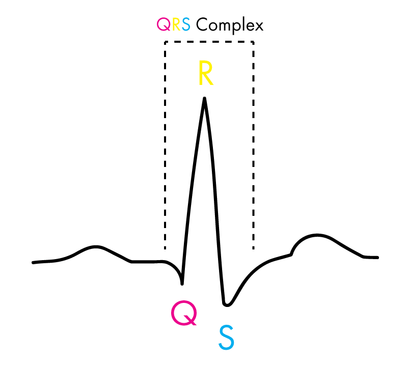
Inter-Beat Interval (IBI). The IBI is the time interval between individual beats of the heart, generally measured in units of milliseconds (ms). Typically, the RR-interval is used for the analysis.
Heart Rate Variability (HRV). HRV expresses the natural variation of IBI values from beat to beat. HRV is closely related to emotional arousal: it has been found to decrease under conditions of acute time pressure and emotional stress (meaning that the heartbeat is more consistent).
HRV has also been found to be significantly reduced in individuals reporting a greater frequency and duration of daily worry [2], as well as in patients suffering from post-traumatic stress disorder (PTSD) [3]. For IBI and HRV analysis, ECG sensors are recommended as they are more sensitive to certain signal characteristics which PPG sensors cannot pick up.
Why combine ECG with other sensors?
Of course, data based on heart rate alone offers valuable insights into nonconscious arousal in response to emotionally-loaded stimuli. However, data solely based on ECG or PPG data can‘t tell us whether the arousal was due to positive or negative stimulus content.
Why? The change in heart rate is in fact identical. Both positive and negative stimuli can result in an increase in arousal triggering changes in heart rate.
In other words: While ECG/PPG are ideal measures to track emotional arousal, they are not able to reveal emotional valence, the direction of an emotion. The true power of ECG/PPG techniques unfolds as these sensors are combined with other data sources such as facial expression analysis, EEG, and eye tracking.
If you’d like to learn more about ECG, and what the heart can tell you about how people feel, download our pocket guide below.
Free 32-page ECG Guide
For Beginners and Intermediates
References
[1] Schacter, S., & Singer, J. (1962). Cognitive, social and physiological determinants of emotional state. Minneapolis: Psychological Review.
[2] Brosschot, J. F., Dijk, E. V., & Thayer, J. F. (2007). Daily worry is related to low heart rate variability during waking and the subsequent nocturnal sleep period. International Journal of Psychophysiology, 63(1), 39-47. doi:10.1016/j.ijpsycho.2006.07.016
[3] Tan, G., Dao, T. K., Farmer, L., Sutherland, R. J., & Gevirtz, R. (2010). Heart Rate Variability (HRV) and Posttraumatic Stress Disorder (PTSD): A Pilot Study. Applied Psychophysiology and Biofeedback, 36(1), 27-35. doi:10.1007/s10484-010-9141-y
Further Reading
Curious to dig deeper into ECG? Have a look at our list of must-reads to learn more about cardiac data recordings and analysis.
- Dutton & Aron (1974). Some evidence for heightened sexual attraction under conditions of high anxiety. Journal of Personality and Social Psychology 30: 510–517. (link)
- Nickel & Nachreiner (2003). Sensitivity and Diagnostics of the 0.1-Hz Component of Heart Rate Variability as an Indicator of Mental Workload. Human Factors 45 (4): 575–590. (link)
- Jönsson (2007). Respiratory sinus arrhythmia as a function of state anxiety in healthy individuals. International Journal of Psycho-physiology 63 (1): 48–54. (link)
- Brosschot, Van Dijk, & Thayer (2007). Daily worry is related to low heart rate variability during waking and the subsequent nocturnal sleep period. International Journal of Psychophysiology 63 (1): 39–47. (link)
- Hagit et al. (1998). Analysis of heart rate variability in posttraumatic stress disorder patients in response to a trauma-related reminder. Biological Psychiatry 44.


