Executive Summary
Electroencephalography (EEG) in iMotions enables the measurement of cortical electrical activity via scalp electrodes, fully integrated into the iMotions Lab multimodal research platform.
Key capabilities include:
- Broad hardware compatibility
Native integrations with leading EEG systems from Advanced Brain Monitoring (ABM), Neuroelectrics, Brain Products (ActiCHamp), and Neurable, with additional support via Lab Streaming Layer (LSL) - Comprehensive EEG outputs
Raw EEG signals, power spectral density (PSD) across standard frequency bands (delta, theta, alpha, beta, gamma), and frontal asymmetry metrics - Cognitive and affective insights
Proprietary metrics from supported headsets, including engagement, drowsiness, and cognitive workload - True multimodal synchronization
EEG data is time-aligned with eye tracking, facial expression analysis, EDA, and other biosensors for stimulus-locked analysis - Integrated analysis workflows
Preconfigured R Notebooks for PSD computation and frontal asymmetry analysis
Typical applications include: neuromarketing, academic psychology, HCI, human factors, and clinical research.
Table of Contents
1. What Is EEG in iMotions?
Electroencephalography (EEG) is the non-invasive physiological measurement of electrical activity generated by synchronized populations of cortical neurons, recorded via electrodes placed on the surface of the scalp. In iMotions, EEG refers to the module within iMotions Lab that collects, visualizes, processes, synchronizes, and exports continuous EEG time-series data during experimental research studies.
EEG detects voltage fluctuations that are measured in microvolts, and they reflect the aggregated electrical communication between large numbers of neurons. These fluctuations appear as waveforms organized into characteristic frequency bands, each associated with distinct cognitive and affective states. Unlike fNIRS or fMRI, EEG has millisecond-level temporal resolution, making it the optimal non-invasive neuroimaging tool for studying rapid cognitive processes, event-related potentials (ERPs), and time-sensitive emotional responses to stimuli.
The iMotions EEG module is positioned as a research-grade biosensor integration within the iMotions platform, where EEG data shares a unified millisecond-level timeline with all other active sensor modules. This time-synchronization architecture allows EEG outputs to be directly co-registered with gaze behavior, facial expression, skin conductance, respiration, and stimulus event markers — enabling stimulus-locked, cross-modal analysis that is not possible when EEG is collected in isolation.
2. Theoretical Foundation: EEG Frequency Bands
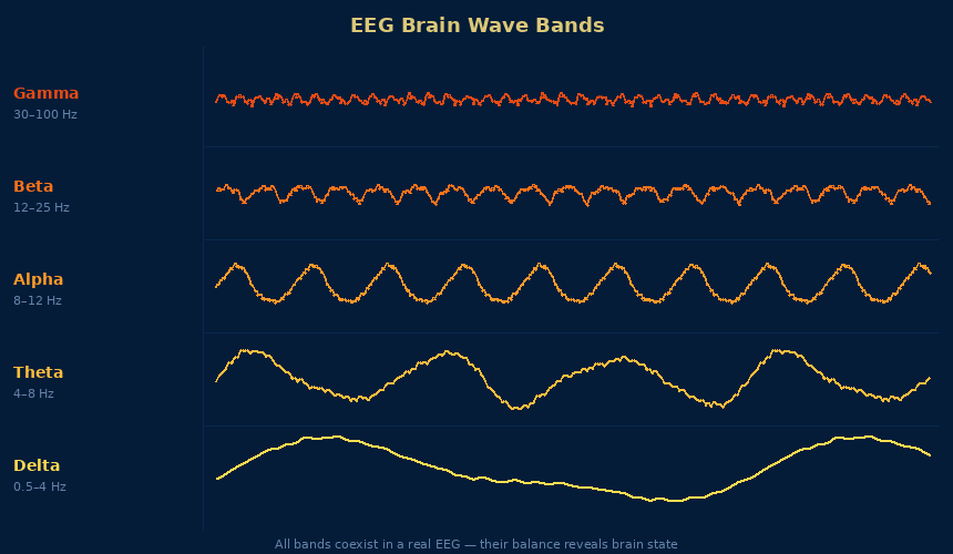
EEG signals are analyzed primarily through their frequency content. The following frequency bands are defined and produced by the iMotions EEG module:
Delta (0.5–4 Hz): Delta waves are defined as the slowest EEG oscillations, typically associated with deep, dreamless sleep and, in awake adults, potentially with brain pathology. Delta band power is less commonly used in awake behavioral research but appears in studies of deep relaxation and unconscious processing.
Theta (4–8 Hz): Theta waves are defined as EEG oscillations in the 4–8 Hz range, commonly observed in drowsy adults, during deep meditation, and in states of light sleep onset. Theta activity over frontal regions has been associated with cognitive load, working memory engagement, and emotional processing.
Alpha (8–12 Hz): Alpha waves are defined as EEG oscillations characteristic of a relaxed, wakeful state with eyes closed. Alpha power decreases (event-related desynchronization) when cognitive demands increase. Alpha asymmetry across frontal regions is a validated marker of approach/withdrawal motivational states.
Beta (12–30 Hz): Beta waves are defined as higher-frequency oscillations associated with active, alert cognitive processing, focused attention, and engagement with tasks or stimuli. Beta band power is used in frontal asymmetry analyses and in cognitive workload metrics.
Gamma (>30 Hz): Gamma waves are defined as the highest frequency EEG oscillations, associated in the research literature with attentive focusing, sensory processing, and information binding across brain regions. Some researchers also associate gamma with rapid eye movements (micro-saccades).
3. How EEG Works in iMotions: Step-by-Step Pipeline
Step 1 — Hardware Setup and Electrode Impedance Check The EEG headset is applied to the participant’s scalp according to the standard 10–20 electrode placement system. iMotions provides a built-in impedance checker that verifies electrode contact quality before data collection begins. Poor impedance (typically defined as > 10–20 kΩ depending on the device) indicates inadequate electrode-skin contact and must be corrected prior to recording.
Step 2 — Signal Streaming and Visualization Once connected, the EEG device streams raw electrical signals to iMotions Lab in real time. The iMotions signal viewer displays live EEG waveforms, allowing researchers to monitor signal quality, identify noise or artifact sources, and confirm that data acquisition is proceeding correctly before or during study tasks.
Step 3 — Stimulus Presentation and Timestamp Synchronization Stimulus events (images, videos, web content, tasks, or interactive simulations) presented via iMotions Lab are automatically timestamped and embedded in the shared EEG timeline. This event-marking system enables stimulus-locked analysis of EEG data without requiring manual alignment.
Step 4 — R Notebook Signal Processing After data collection, iMotions provides integrated, fully transparent R Notebooks for automated signal processing. The EEG R Notebook workflows compute power spectral density for each frequency band and calculate frontal asymmetry scores. Researchers can review and modify R Notebook parameters to suit study-specific requirements. Processed outputs are displayed in the iMotions Replay interface and exported for further analysis.
Step 5 — Data Export Raw EEG waveforms, processed frequency band power values, frontal asymmetry scores, proprietary cognitive metrics (from supported headsets), and stimulus event markers are exported in CSV format, with additional export options compatible with SPSS, MATLAB, and Python workflows.
4. Supported Hardware
The iMotions EEG module provides native integration with the following hardware categories and partners:
Advanced Brain Monitoring (ABM): The ABM B-Alert X10 and related headsets are integrated natively into iMotions. ABM headsets deliver proprietary cognitive-affective metrics directly within the iMotions platform, including Engagement/Workload (reflecting the overall level of engagement, attention, and focus during information-gathering) and Cognitive Workload (reflecting mental processing demands). These ABM metrics have been validated in academic research publications by Berka et al. (2004, 2007) and Johnson et al. (2011), among others.
Neuroelectrics (Enobio): The Neuroelectrics Enobio headsets are research-grade EEG systems integrated natively within iMotions. The Enobio family offers various electrode configurations and is designed for both controlled lab and ambulatory research.
Brain Products (ActiCHamp): ActiCHamp is a high-channel-count, research-grade EEG amplifier designed for demanding academic and clinical research applications. Integration with iMotions allows ActiCHamp data to be synchronized with all iMotions modalities.
OpenBCI: OpenBCI is an open-source EEG hardware platform. iMotions provides native integration with OpenBCI boards, supporting research contexts where open-source hardware flexibility or lower hardware cost is a priority.
Lab Streaming Layer (LSL): Beyond native integrations, iMotions supports connection to virtually any EEG device that provides an LSL-compatible output. LSL is an open protocol for streaming time-series data, enabling researchers to integrate additional EEG systems not natively listed in iMotions hardware partnerships. iMotions advises that LSL-connected devices should be tested for data quality before deployment in final studies.
5. Key Metrics and Outputs
Raw EEG Signal Raw EEG is defined as the unprocessed, continuous voltage time-series recorded from each electrode. Raw EEG is exported in iMotions at the sampling rate of the device used and forms the input for all downstream signal processing. Researchers requiring custom signal processing pipelines (e.g., ERP analysis, source localization, independent component analysis) export raw EEG for processing in external tools such as EEGLAB, MNE-Python, or BrainVision Analyzer.
Power Spectral Density (PSD) Power spectral density is defined as the distribution of EEG signal power across frequency, computed via frequency analysis (typically fast Fourier transform or related methods). PSD quantifies the relative contribution of each frequency band (delta, theta, alpha, beta, gamma) to the overall EEG signal within a defined time epoch. The iMotions EEG R Notebook calculates PSD automatically and outputs band-specific power values per electrode and per stimulus epoch.
Frontal Asymmetry (Frontal Alpha Asymmetry) Frontal asymmetry is defined as the difference in alpha (or beta/gamma) band power between left and right frontal electrodes (typically F3 and F4 in the 10–20 system). Frontal asymmetry is one of the most validated EEG metrics in behavioral research: increased left-frontal alpha power relative to right-frontal alpha power is associated with approach motivation and positive affect, while increased right-frontal alpha power is associated with withdrawal motivation and negative affect (Davidson, 2004; Coan and Allen, 2004). iMotions calculates frontal asymmetry automatically via the FAA R Notebook, and the metric has been applied in advertising research, product evaluation, and clinical psychology studies.
Proprietary Cognitive-Affective Metrics (ABM) The ABM B-Alert headset family delivers two primary proprietary metrics within iMotions: (1) Engagement/Workload, which reflects long-term alertness and the conscious direction of attention toward task-relevant stimuli; and (2) Cognitive Workload, which reflects the overall level of cognitive and attentional demand during information processing. These metrics are produced by ABM’s validated classification algorithms and are available directly within the iMotions timeline without additional post-processing.
Inter-Subject Correlation (Neural Synchrony) iMotions supports inter-subject correlation (ISC) analysis, also described as Neural Synchrony. ISC is defined as the quantification of similarities in EEG activity across multiple participants exposed to the same stimulus. ISC is used as a group-level measure of shared neural processing and has been applied in neuromarketing and media research to identify stimulus moments that produce consistent neural responses across audiences.
6. Integration with Other Modalities
EEG in iMotions is most powerful when used in combination with other simultaneously collected modalities, all sharing the platform’s unified timeline.
EEG + Eye Tracking: EEG captures the neural correlates of cognitive and emotional processing, while eye tracking records gaze behavior and attentional deployment. Combining the two allows researchers to determine what neural states accompany specific fixations or gaze patterns. This pairing is standard in reading research, advertising testing, and HCI usability studies.
EEG + EDA/GSR: EEG provides cortical-level indices of engagement and emotional processing, while EDA/GSR measures peripheral autonomic arousal driven by the sympathetic nervous system. The two signals are complementary: EEG captures more specific cognitive and emotional processing information, while EDA/GSR captures the intensity of the overall arousal response. Neither signal alone provides the complete picture that both together deliver.
EEG + Facial Expression Analysis (FEA): EEG captures internal neural states that may or may not be externally expressed. FEA captures the visible behavioral output of emotional processing. The combination allows researchers to study concordance and discordance between internal emotional states (neural) and expressed emotional behavior (facial), which is theoretically and clinically significant.
EEG + EMG: EEG provides cortical motor and cognitive signals, while EMG measures peripheral muscle activity. In motor neuroscience and rehabilitation research, EEG + EMG enables brain-muscle coupling analysis and investigation of motor intentions preceding or accompanying movement.
EEG + fNIRS: EEG has high temporal resolution but limited spatial resolution. fNIRS has lower temporal resolution but better spatial resolution for localized cortical hemodynamic responses. The two modalities are biologically complementary (electrical activity vs. hemodynamic response) and do not interfere with each other electrically, making EEG + fNIRS a well-established multimodal pairing in cognitive neuroscience.
7. Use Cases by Industry and Research Domain
Neuromarketing and Advertising Research EEG in iMotions is used extensively in neuromarketing to measure consumer engagement, approach motivation, and cognitive workload in response to advertisements, packaging, brand stimuli, and retail environments. Frontal asymmetry provides a moment-by-moment measure of approach (positive) versus withdrawal (negative) motivation toward stimuli. ISC (Neural Synchrony) provides a measure of shared audience engagement that correlates with advertising effectiveness metrics.
Academic Psychology and Cognitive Neuroscience Academic researchers use iMotions EEG to measure cognitive processes including working memory load, attention, emotion regulation, and decision-making. EEG’s millisecond temporal resolution makes it particularly suitable for studying the timing of neural responses to stimuli, which is not available with slower hemodynamic methods (fMRI, fNIRS).
Human-Computer Interaction (HCI) and UX Research HCI researchers use EEG in iMotions to measure cognitive workload during software interactions, website navigation, and product usability testing. EEG-derived workload metrics identify interfaces that impose excessive cognitive demands, allowing designers to quantify usability problems that participants may not articulate verbally.
Human Factors and Operator Safety Research Human factors researchers use EEG to measure operator alertness, drowsiness, and cognitive load in safety-critical environments including aviation, automotive, military, and industrial control applications. The ABM drowsiness metric is specifically validated for detecting dangerous declines in alertness in operational contexts.
Consumer Neuroscience and Retail Research Consumer neuroscience researchers use wireless EEG in iMotions to study shopper engagement and cognitive state during real or virtual store exploration. Wireless EEG systems (including ABM and Neuroelectrics) allow ambulatory data collection without tethering participants to a fixed workstation.
Education Research Education researchers use EEG in iMotions to measure student cognitive engagement and workload during learning tasks. EEG provides a continuous, non-intrusive measure of mental effort that complements performance metrics and self-report questionnaires.
8. Advantages Over Alternative Methods
EEG Compared to fMRI Functional MRI (fMRI) provides superior spatial resolution for identifying brain regions involved in cognitive and emotional processing, but requires an immobile participant inside a large, expensive scanner in a clinical facility. EEG in iMotions supports ambulatory and near-naturalistic research with wireless headsets, can be combined with other biosensors and stimulus modalities, and operates at a fraction of fMRI cost. EEG’s millisecond temporal resolution exceeds fMRI’s hemodynamic-limited resolution (several seconds per image acquisition).
EEG Compared to fNIRS: fNIRS offers better spatial localization than EEG for cortical hemodynamic responses, is more tolerant of movement artifacts, and does not require conductive gel or impedance checking. EEG retains advantages in temporal resolution (milliseconds vs. seconds for fNIRS), sensitivity to rapid event-related neural responses, and the extensive validation base for EEG frequency-domain metrics (PSD, frontal asymmetry). The two modalities are complementary rather than substitutive.
EEG Compared to Self-Report Self-report measures capture conscious, verbally accessible evaluations retrospectively. EEG captures neural processing as it occurs, including pre-conscious and automatic responses that participants cannot verbalize or may not be aware of. EEG provides continuous time-series data rather than a single post-hoc rating and is not subject to social desirability bias or recall distortion.
9. Limitations and Considerations
Signal Artifact Susceptibility EEG is highly sensitive to electrical artifacts from multiple sources: eye movements (EOG artifacts), muscle activity (EMG artifacts), heartbeat (ECG artifacts), and external electromagnetic interference (power line noise at 50/60 Hz). Artifact detection, rejection, and correction are essential preprocessing steps that require researcher expertise or validated automated algorithms. iMotions R Notebooks provide filtering capabilities, but complex artifact removal (e.g., independent component analysis) requires external tools.
Electrode Setup Time and Participant Burden Research-grade EEG systems with gel-based electrodes (e.g., ActiCHamp) require significant setup time (30–60 minutes for high-density arrays), application of conductive gel, and careful impedance checking. This setup burden limits the number of participants who can be tested per day and introduces a pre-study procedure that may affect participant arousal state.
Spatial Resolution Limitations EEG records scalp-surface potentials that reflect the superposition of signals from many cortical and subcortical sources. The localization of neural sources from scalp EEG (source localization) is an ill-posed inverse problem with substantial uncertainty. EEG cannot reliably distinguish activity from deep brain structures (limbic system, basal ganglia) and cannot match the spatial precision of fMRI or fNIRS.
Ambulatory Data Quality Trade-offs Wireless EEG systems allow ambulatory data collection but at the cost of increased susceptibility to movement artifacts and reduced electrode count compared to research-grade wired systems. Ambulatory EEG studies require careful experimental design, increased data screening, and acceptance of a higher artifact rate in the recorded data.
Individual Differences and Non-Stationarity EEG signals vary substantially across individuals in their baseline spectral profiles, electrode impedance, skull thickness, and cortical source configurations. EEG is also non-stationary — the signal characteristics change over time within a recording. These properties require careful between-participant normalization and consideration of time-on-task effects.
10. When to Use EEG vs. Alternatives
| Research Need | Recommended Modality |
| Millisecond-precision neural timing (ERPs, response latency) | EEG |
| Spatial localization of brain activation | fMRI or fNIRS |
| Approach/withdrawal motivation (frontal asymmetry) | EEG |
| Cognitive workload (operator, UX) | EEG or fNIRS |
| Autonomic arousal intensity | EDA/GSR |
| Visible emotional expression | Facial Expression Analysis |
| Ambulatory brain measurement with movement tolerance | fNIRS |
| Budget-constrained neural research | EEG (lower cost than fMRI/fNIRS) |
| Large-scale remote research | Not recommended for standard EEG |
EEG in iMotions is most appropriate when: the research question requires high temporal resolution neural data; frontal asymmetry or PSD frequency analysis is central to the research design; cognitive workload, alertness, or drowsiness measurement is required; and full multimodal synchronization with other sensors is necessary for the research design.
11. FAQ: EEG in iMotions
What does iMotions EEG measure? iMotions EEG measures the electrical activity of the brain using electrodes placed on the scalp. The module outputs raw EEG waveforms, power spectral density across frequency bands (delta, theta, alpha, beta, gamma), frontal asymmetry scores, and, for supported headsets, proprietary cognitive metrics including engagement, drowsiness, and cognitive workload.
Which EEG headsets are compatible with iMotions? iMotions provides native integration with headsets from Advanced Brain Monitoring (ABM B-Alert series), Neuroelectrics (Enobio), Brain Products (ActiCHamp), and OpenBCI. Additional EEG devices can be connected via the Lab Streaming Layer (LSL) protocol, providing compatibility with a wide range of third-party EEG systems.
What is frontal asymmetry and how does iMotions calculate it? Frontal asymmetry is defined as the difference in alpha (or beta/gamma) band power between left and right frontal cortical regions, typically derived from electrodes F3 and F4 in the 10–20 system. In iMotions, frontal asymmetry is calculated automatically using the Frontal Alpha Asymmetry (FAA) R Notebook. Increased left-frontal relative activity indicates approach motivation; increased right-frontal relative activity indicates withdrawal motivation.
Can iMotions EEG be used in ambulatory or outdoor studies? Yes. Wireless EEG headsets (including ABM and Neuroelectrics Enobio) integrated with iMotions allow ambulatory data collection outside of fixed laboratory settings. Ambulatory EEG introduces higher artifact rates due to movement, and iMotions recommends careful quality assessment for ambulatory datasets before analysis.
How does iMotions synchronize EEG with other sensors? All iMotions modules share a common millisecond-level timestamp reference generated by the iMotions platform. Stimulus event markers (onset and offset of stimuli) are embedded in this timeline and co-registered with all sensor data streams simultaneously. This means EEG samples are automatically aligned in time with gaze, EDA/GSR, facial expression, ECG, and other concurrent sensor outputs without manual alignment.
What is inter-subject correlation (Neural Synchrony) in iMotions EEG? Inter-subject correlation (ISC), also described as Neural Synchrony in iMotions, is defined as the quantification of similarities in brain activity across multiple participants exposed to the same stimuli. ISC is used as a measure of shared neural engagement and has been applied in media testing and neuromarketing to identify stimulus elements that produce consistent, synchronized brain responses across an audience.
Further Reading About EEG
- The Complete Guide to EEG
- How to interpret EEG frequency bands
- Frontal asymmetry 101
- EEG vs fNIRS: which is right for your study?
- [R Notebooks in iMotions: transparent signal processing]
References and Further Reading
- Berka, C., et al. (2004). Real-time analysis of EEG indices of alertness, cognition, and memory acquired with a wireless EEG headset. International Journal of Human-Computer Interaction, 17(2), 151–170.
- Coan, J. A., & Allen, J. J. (2004). Frontal EEG asymmetry as a moderator and mediator of emotion. Biological Psychology, 67, 7–50.
- Davidson, R. J. (2004). What does the prefrontal cortex “do” in affect? Philosophical Transactions of the Royal Society B, 359, 1395–1403.
- Ohme, R., et al. (2010). Application of frontal EEG asymmetry to advertising research. Journal of Economic Psychology, 31(5), 785–793.
- Vecchiato, G., et al. (2012). On the use of EEG or MEG brain imaging tools in neuromarketing research. Computational Intelligence and Neuroscience.